
Plik:Oncocytoma of the Salivary Gland.jpg
Oncocytoma_of_the_Salivary_Gland.jpg (550 × 335 pikseli, rozmiar pliku: 25 KB, typ MIME: image/jpeg)
| Plik Oncocytoma of the Salivary Gland.jpg znajduje się w Wikimedia Commons – repozytorium wolnych zasobów. Dane z jego strony opisu znajdują się poniżej. |
Opis
| Opis |
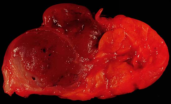
Oncocytoma of the Salivary Gland This lesion presented as a lateral anterior neck mass. At surgery, it was found to be a soft 3.0 x 2.1 x 1.8 cm tumor of the submandibular salivary gland. The photo shows the characteristic dark color of an oncocytoma, a rare type of benign neoplasm, at the left side of the image (the normal lobulated salivary gland tissue is to the right). Excision is curative. Since this specimen was photographed in the fresh state, it is various shades of red due to blood staining. A little formalin fixation would be expected to better emphasize the color difference between the tumor and the normal gland tissue. The photo was shot with a Minolta X-370 with 100mm Rokkor bellows lens on Kodak Elite ISO 100 daylight-balanced transparency film. I used a blue filter to compensate for the tungsten illumination. Photograph by Ed Uthman, MD. Public domain. Posted 19 May 00 |
| Źródło | http://web2.airmail.net/uthman/specimens/index.html |
| Autor | |
| Licencja (Ponowne użycie tego pliku) |
PD |
Licencja
| Ten utwór został udostępniony jako własność publiczna przez jego autora, Ed Uthman. Dotyczy to całego świata. W niektórych krajach może nie być to prawnie możliwe, jeśli tak, to: Ed Uthman zapewnia każdemu prawo do użycia tej pracy w dowolnym celu, bez żadnych ograniczeń, chyba że te ograniczenia są wymagane przez prawo.
|
Historia pliku
Kliknij na datę/czas, aby zobaczyć, jak plik wyglądał w tym czasie.
| Data i czas | Miniatura | Wymiary | Użytkownik | Opis | |
|---|---|---|---|---|---|
| aktualny | 11:53, 5 cze 2006 | | 550 × 335 (25 KB) | Patho | {{Information| |Description=Oncocytoma of the Salivary Gland This lesion presented as a lateral anterior neck mass. At surgery, it was found to be a soft 3.0 x 2.1 x 1.8 cm tumor of the submandibular salivary gland. The photo shows the characteristic dar |
Lokalne wykorzystanie pliku
Poniższa strona korzysta z tego pliku:
Globalne wykorzystanie pliku
Ten plik jest wykorzystywany także w innych projektach wiki:
- Wykorzystanie na de.wikipedia.org
- Wykorzystanie na de.wikibooks.org
- Wykorzystanie na en.wikipedia.org
- Wykorzystanie na fr.wikipedia.org